Poor Piggy: Pig Dissection Lab
In replace of a video, I decided to write about it instead of show it.

External Anatomy:
Usually, the fetal pig is a light beige/ off pink color. To dissect it, you must tie down the wrists on the plate to have the ventral area exposed for dissection. The wrists of the pig is cut out of the diagram, but just like humans, it would be located near the ends of the arm. The shoulder is spread openly, which can be found after the elbow of the arm. Digits are essentially the fingers and toes of a pig. They are connected to the wrist, at the end of the arm. Those two structures allow the pig to move. The thoracic cavity is everything from the diaphragm and up. We'll talk about what goes into that portion of the pig later. The abdominal cavity should be everything below the thoracic cavity. To determine the sex, you look at the urogenital opening. On males, it should be located near the umbilical cord. For females, the opening is near the anus.
Digestive System:
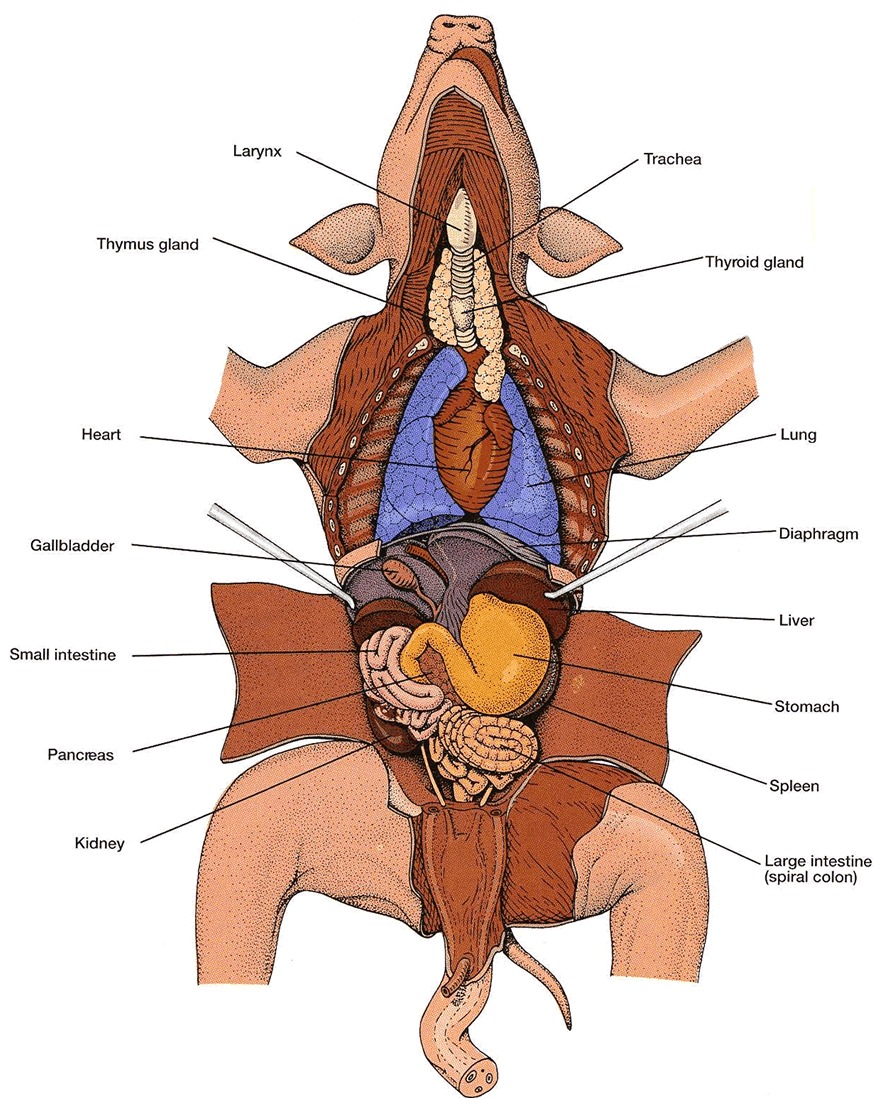
In a digestive system, you have to start with the chewing of the food, and that is where the mouth is. The hard palates, which are the more tough area of the mouth, are on the roof of the mouth and is connected to the esophagus, right behind the trachea. It helps the pig eat and chew the food. The soft palates are further in the mouth, helping the already chewed food be digested easier. We move down the pig and find the more obvious structures. The stomach, a large, yellow circle depicted in the diagram, is the second part of digestion (first part being the intake of food through the esophagus). It contains enzymes that help with the process. Going through the flow of food, next would be the liver. It is above the stomach and produced bile to aid with digestion. The pancreas is intertwining with the end of the sack of the stomach. It secretes enzymes to the small intestine, which isn't so small. It sits next to the stomach and pancreas, and is where most of the digesting and absorbing of nutrients occur. The smaller intestine, which is ironically called the large intestine is below the stomach and is used to help pass the ingested food that can't be broken down from the body. The reason why there isn't any food in the stomach and intestines is because it is still a fetal pig that was killed before being able to eat anything. Lastly, there is the rectum. It is a tube, leading to the anus, and is the end of the large intestine. It is the last, internal part of the digestive system that hold the feces before being pushed out of the anus.
Respiratory System:
What is needed to breathe? Lungs, diaphragm, and trachea. The lungs are surrounding the diaphragm and rib cage in the thoracic cavity, that exchanges gas, releasing carbon dioxide. They have the texture of a sponge and are a dark red color. The diaphragm is the thin muscle below the lungs. It divides the thoracic and abdominal cavity. It pulls in air into the lungs. The trachea is the white, tube-like organ. It carries oxygen to the lungs. Even by just looking at it, you can tell it's rigid and bony.

Circulatory System:
In the circulatory system, the main organ is the heart. The heart is located in between the lungs and is centered of the body. This way, it is easier to transport blood through the blood vessels to the different parts of the body. There are four chambers: right auricle, left auricle, right ventricle, left ventricle, and right ventricle. From the labeled diagram, you can locate the aorta, which oxygenates blood to the tissues. It't the largest blood vessel. The coronary artery is the blood vessel that supplies blood to the heart muscle. It also separates the two ventricles. Next, the superior vena cava carries de-oxygen from the upper part of the body to the right atrium, while the inferior vena cava carries de-oxygen from the lower part of the body to the right atrium. It is locate to the aorta. The renal artery and vein are next to the kidney. The arteries' function supplies oxygen blood to the kidney, and the veins remove the de-oxygenized blood from the kidney. The circulatory system is very important to mammals because its mandatory for them to have blood and oxygen flow, that is regulated, in their body. It keeps all the other systems in check. Another point would be it supplies the mammal with nutrients to maintain their life.
Urinary and Reproductive System:
The kidney is hidden behind the large intestines and excretes waste and water from the body. If the kidney stops working, the excess waste would be contained inside and your body would be filled with all that bad stuff. When a kidney fails, this is called uremia. Ureter is next, which is a thick-walled tube that carries waste from the kidney to the urinary bladder. The bladder is located near the reproductive areas of the fetal pig. It stores all the waste before excreting.
Endocrine System:
The purpose of the endocrine system is to have a longer lasting control of the bodily functions. In the endocrine system, there are two main organs: the thyroid and spleen. The thyroid is located on the trachea and produces hormones and regulates metabolism. The spleen is near the stomach and produces, stores, and eliminates red blood cells.
Relate and Review:
The dissection would have been a great visual representation of all of the functions and locations of the organs and such. It's like looking at a map while learning world geography to relate to what you are learning. I actually did not do the dissection so I do not have a favorite part, although looking at the pictures of the structures was interesting. I felt that if I did do the dissection, I would feel stronger about my knowledge of the body structures. Additionally, I would feel stronger as a person for doing something that I am uncomfortable doing. On the other hand, my parents did highly suggest that I skip this lab because of our religion.
External Anatomy:
Usually, the fetal pig is a light beige/ off pink color. To dissect it, you must tie down the wrists on the plate to have the ventral area exposed for dissection. The wrists of the pig is cut out of the diagram, but just like humans, it would be located near the ends of the arm. The shoulder is spread openly, which can be found after the elbow of the arm. Digits are essentially the fingers and toes of a pig. They are connected to the wrist, at the end of the arm. Those two structures allow the pig to move. The thoracic cavity is everything from the diaphragm and up. We'll talk about what goes into that portion of the pig later. The abdominal cavity should be everything below the thoracic cavity. To determine the sex, you look at the urogenital opening. On males, it should be located near the umbilical cord. For females, the opening is near the anus.
Digestive System:
In a digestive system, you have to start with the chewing of the food, and that is where the mouth is. The hard palates, which are the more tough area of the mouth, are on the roof of the mouth and is connected to the esophagus, right behind the trachea. It helps the pig eat and chew the food. The soft palates are further in the mouth, helping the already chewed food be digested easier. We move down the pig and find the more obvious structures. The stomach, a large, yellow circle depicted in the diagram, is the second part of digestion (first part being the intake of food through the esophagus). It contains enzymes that help with the process. Going through the flow of food, next would be the liver. It is above the stomach and produced bile to aid with digestion. The pancreas is intertwining with the end of the sack of the stomach. It secretes enzymes to the small intestine, which isn't so small. It sits next to the stomach and pancreas, and is where most of the digesting and absorbing of nutrients occur. The smaller intestine, which is ironically called the large intestine is below the stomach and is used to help pass the ingested food that can't be broken down from the body. The reason why there isn't any food in the stomach and intestines is because it is still a fetal pig that was killed before being able to eat anything. Lastly, there is the rectum. It is a tube, leading to the anus, and is the end of the large intestine. It is the last, internal part of the digestive system that hold the feces before being pushed out of the anus.
Respiratory System:
What is needed to breathe? Lungs, diaphragm, and trachea. The lungs are surrounding the diaphragm and rib cage in the thoracic cavity, that exchanges gas, releasing carbon dioxide. They have the texture of a sponge and are a dark red color. The diaphragm is the thin muscle below the lungs. It divides the thoracic and abdominal cavity. It pulls in air into the lungs. The trachea is the white, tube-like organ. It carries oxygen to the lungs. Even by just looking at it, you can tell it's rigid and bony.
Circulatory System:
In the circulatory system, the main organ is the heart. The heart is located in between the lungs and is centered of the body. This way, it is easier to transport blood through the blood vessels to the different parts of the body. There are four chambers: right auricle, left auricle, right ventricle, left ventricle, and right ventricle. From the labeled diagram, you can locate the aorta, which oxygenates blood to the tissues. It't the largest blood vessel. The coronary artery is the blood vessel that supplies blood to the heart muscle. It also separates the two ventricles. Next, the superior vena cava carries de-oxygen from the upper part of the body to the right atrium, while the inferior vena cava carries de-oxygen from the lower part of the body to the right atrium. It is locate to the aorta. The renal artery and vein are next to the kidney. The arteries' function supplies oxygen blood to the kidney, and the veins remove the de-oxygenized blood from the kidney. The circulatory system is very important to mammals because its mandatory for them to have blood and oxygen flow, that is regulated, in their body. It keeps all the other systems in check. Another point would be it supplies the mammal with nutrients to maintain their life.
Urinary and Reproductive System:
The kidney is hidden behind the large intestines and excretes waste and water from the body. If the kidney stops working, the excess waste would be contained inside and your body would be filled with all that bad stuff. When a kidney fails, this is called uremia. Ureter is next, which is a thick-walled tube that carries waste from the kidney to the urinary bladder. The bladder is located near the reproductive areas of the fetal pig. It stores all the waste before excreting.
Endocrine System:
The purpose of the endocrine system is to have a longer lasting control of the bodily functions. In the endocrine system, there are two main organs: the thyroid and spleen. The thyroid is located on the trachea and produces hormones and regulates metabolism. The spleen is near the stomach and produces, stores, and eliminates red blood cells.
Relate and Review:
The dissection would have been a great visual representation of all of the functions and locations of the organs and such. It's like looking at a map while learning world geography to relate to what you are learning. I actually did not do the dissection so I do not have a favorite part, although looking at the pictures of the structures was interesting. I felt that if I did do the dissection, I would feel stronger about my knowledge of the body structures. Additionally, I would feel stronger as a person for doing something that I am uncomfortable doing. On the other hand, my parents did highly suggest that I skip this lab because of our religion.


Comments
Post a Comment